World Glaucoma Week: Symptoms and Treatment: Latest advance techniques in India
Like | DoctorBabu | March 13, 2018 | Health eyecare, glaucoma, health, healthy tips
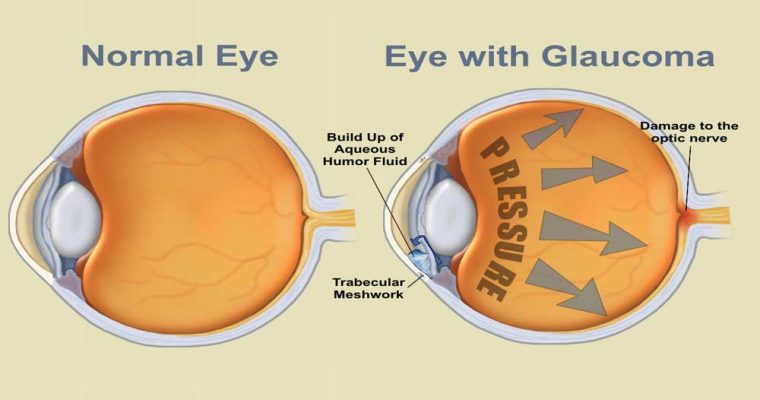
According to Glaucoma Society of India, glaucoma is the third leading cause of blindness in India. Glaucoma shows no symptoms, and there is no way you can prevent it. Glaucoma is a disease in which the optic nerve, the nerve that connects the eye to the brain, is damaged. Generally, this damage is caused by high pressure in the eye. The eye makes and drains fluid when the drainage system becomes impaired pressure from the excess fluid builds up in the eye and results in glaucoma. It is a progressive condition that, without treatment, will lead to loss of peripheral (side) vision and, ultimately, blindness.
Diagnosis
Glaucoma is diagnosed by measuring the eye pressure and estimating the damage to the optic nerve. Dr Nitin Deshpande, Glaucoma Consultant, says that glaucoma can be diagnosed in two ways.
1. Perimetry–This test maps your complete field of vision and is subjective to the patient’s response. In this test, you will be given the controls to a machine, and a light is flashed in your peripheral vision., With the help of the buttons on the machine, you will have to respond to the light when it is flashed. This will help your doctor map your entire field of peripheral vision. This test helps determine the functioning of your eye.
The test gives result in a very short period. However, it is not very accurate as it also depends on how well the patient responds to the flashed light.
New software like Swedish Interactive Threshold Algorithm (SITA) is coupled with Perimetry to help doctors determine the loss of vision more accurately. This software is highly sensitive and makes the test more patient friendly giving the results in a very short period.
2. Optical Coherence Tomography–This is a non-invasive diagnostic tool that uses infrared light to map different layers of the retina and helps the doctor in measuring the thickness of these layers. This tool can help your doctor predict your risk of glaucoma seven to eight years in advance.
Latest techniques used to treat glaucoma.
usually aims at preventing further damage to the optic nerve and prevent loss of vision. Glaucoma has been traditionally treated with eye drops and surgery which help in reducing the eye pressure and help in draining the built-up fluid.
Non-invasive methods
1. Trabeculoplasty– Laser trabeculoplasty (truh-BEK-u-low-plas-tee) is an option for people with open-angle glaucoma. It’s done in your doctor’s office. He or she uses a laser beam to open clogged channels in the trabecular meshwork. It may take a few weeks before the full effect of this procedure becomes apparent.
Surgical interventions
1. Trabeculectomy– It is the gold standard in glaucoma treatment and was invented 30 years ago, but is still very effective. It creates a guarded pathway by removing a piece of tissue in the drainage angle of the eye. This pathway helps to drain out the fluid and thereby maintain the eye pressure.
To prevent the failure of this technique, it is modified with implants like:.
a. Ex-Press Glaucoma Shunt–The express shunt is a micro tube that prevents the closing of the channel that is created to drain the fluid.
b. Collagen implants–This implant too prevents the closure of the channel along with preventing fibrosis. All these techniques are a one-day procedure.
2. Visco Canalostomy–It is a minimally invasive glaucoma surgery (MIGS) where the channels in the eye are opened up using a high viscosity solution such as sodium hyaluronate and fluid is drained out to reduce the pressure in the eye.
3. Ahmed Glaucoma Valve surgery–It is one of the most common valves that is used especially when the intraocular pressure in high and cannot be controlled with the help of eye drops. This valve is a tube that drains the fluid out of the eye.
Post-operative care–Dr Deshpande says that patients should use the prescribed eye drops and follow up regularly with the doctor. The key is to prevent the eye from infections.
Leave a Reply