Eyes: Introduction and how they work
Like | DoctorBabu | January 15, 2018 | Health eye, health, healthy tips, retina
Eyes are special sense organ capable of receiving visual images, which are then carried to the brain.
The human eye belongs to a general group of eyes found in nature called “camera-type eyes.” Just as a camera lens focuses light onto film, a structure in the eye called the cornea focuses light onto a light-sensitive membrane called the retina.
Eye health is an important part of overall health. If any part of your visual system is not working, or not conveying the appropriate messages to your brain, vision suffers. Our eyes allow us to appreciate the beauty of the world, experience the joy of learning new activities, and undertake new adventures. Knowing the anatomy of your eyes and having regular exams is the best way to keep your eyes healthy and your vision intact.
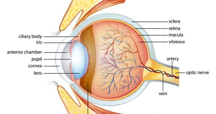
Anatomy of the eye
The tissues of the eye can be split into three types:
Refracting tissues that focus light
Light-sensitive tissues
Support tissues
Refracting tissues
Refracting tissues focus incoming light onto the light-sensitive tissues, to give us a clear, sharp image. If they are the wrong shape, misaligned, or damaged, vision can be blurry.
The refracting tissues include:
The pupil is the hole located in the center of the iris. It allows light to enter the eye. The pupil appears black because light rays entering the pupil are absorbed by the tissues inside the eye. Or they are absorbed after diffused reflections within the eye.
The iris is a thin, circular structure in the eye. It controls the diameter and size of the pupil. The color of the iris is often referred to as eye color.
The cornea is the transparent front part of the eye that covers the iris, pupil, and anterior chamber. The cornea with the anterior chamber and lens refracts light with the cornea. This accounts for approximately two-thirds of the eye’s total optical power.
The crystalline lens is a transparent and biconvex structure. Along with the cornea, it helps to refract light to focus on the retina. By changing shape, the lens functions to change the focal distance of the eye. This happens so that it can focus on objects at various distances.
Two fluids circulate throughout the eyes to provide structure and nutrients. These fluids are:
Vitreous fluid: Found in the back section of the eye, vitreous fluid is thick and gel-like. It makes up the majority of the eye’s mass.
Aqueous fluid: This is more watery than vitreous fluid and circulates through the front of the eye.
Light-sensitive tissues: Retina
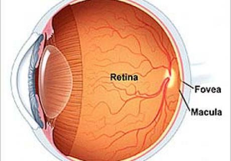
The retina is a light-sensitive layer of tissue lining the surface of the eye. It captures light sent through the cornea and crystalline lens. It then creates an image by triggering nerve impulses that pass to various visual centers of the brain via the optic nerve.
The macula and fovea are small areas within the retina that contain the rods and cones. These structures determine the color and shape of the image you are viewing.
Support tissues
The sclera (the white part of the eye) is the opaque, fibrous, protective outer layer.
Conjunctiva: A thin, transparent membrane that covers most of the white of the eye, and the inside of the eyelids. It helps lubricate the eye and protect it from microbes.
Choroid: A layer of connective tissue between the retina and sclera. It contains a high concentration of blood vessels. It is just 0.5 mm thick and contains light-absorbing pigment cells that help reduce reflections in the retina.
Eye conditions
As with any part of the body, problems with our sight can arise from illness, injury, or age. Below are just some of the conditions that can affect the eyes:
Age-related macular degeneration: Age-related macular degeneration (AMD) is the physical disturbance of the center of the retina called the macula.
Amblyopia: This begins in childhood and is often called lazy eye. One eye does not develop properly because the other, stronger eye dominates.
Anisocoria: This occurs when pupils are an unequal size. It can be a harmless condition or a symptom of a more serious medical problem.
Astigmatism: The cornea or lens is incorrectly curved so that light is not focused properly on the retina.
Cataracts: Cataracts are a degenerative form of eye disease in which the lens gradually becomes opaque and vision mists over.

Colorblindness: Colour blindness is not actually blindness in the true sense but rather is a colour vision deficiency—people who are affected by it simply do not agree with most other people about colour matching.
Conjunctivitis or pink eye: This is a common infection of the conjunctiva, which covers the front of the eyeball.
Detached retina: A condition when the retina comes loose. It requires urgent treatment.
Diplopia or double vision: This can be caused by several conditions that are often serious and should be checked by a doctor, as soon as possible.
Floaters: These are specks that drift across a person’s visual field. They are normal but can also be the sign of something more serious, such as retinal detachment.
Glaucoma: Glaucoma occurs when a build-up of fluid in the eye creates pressure, damaging the optic nerve.
Myopia: This is otherwise known as nearsightedness. With myopia, it is difficult to see things that are far away.
Optic neuritis: The optic nerve becomes inflamed, often due to an overactive immune system.
Strabismus: The eyes point in different directions; it is particularly common among children.
Leave a Reply